問題詳情
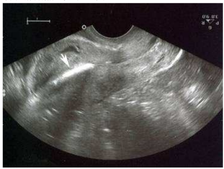
29.一位26歲生過三胎之年輕婦女因不正常陰道出血而就醫,其婦科史有裝置子宮避孕器三年。理學檢查經陰道診視發現子宮頸表面平滑,血液自子宮頸口處流出,子宮頸口未見有子宮避 孕器之尾巴線;觸診發現子宮大小正常且無觸壓痛之情形。經陰道超音波掃描子宮之情形如 下圖,箭頭所指之最適合診斷為:
(A)子宮內膜增殖(endometrial hyperplasia)
(B)子宮內膜息肉(endometrial polyp)
(C)子宮腔內血腫(hematometra)
(D)子宮內避孕器(intrauterine device)
(A)子宮內膜增殖(endometrial hyperplasia)
(B)子宮內膜息肉(endometrial polyp)
(C)子宮腔內血腫(hematometra)
(D)子宮內避孕器(intrauterine device)
參考答案
答案:D
難度:簡單0.857143
統計:A(2),B(0),C(1),D(24),E(0)
難度:簡單0.857143
統計:A(2),B(0),C(1),D(24),E(0)
用户評論
【霸氣阿宏】評論
Fig. 3.Transvaginal ultrasonographic appearance of T-shaped intrauterine devices (IUDs).A, B. Two-dimensional (2D) sagittal (A) and transverse (B) sonograms show hyperechoic levonorgestrel-releasing IUD in the endometrial cavity. C, D. 2D sagittal (C) and transverse (D) sonograms show the bright echo of the copper IUD with marked posterior shadowing. E. Three-dimensional coronal reformatted sonogram demonstrates the properly positioned copper IUD within the endometrial cavity (arrows)...